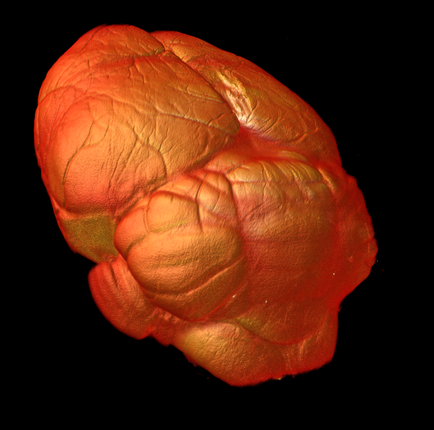
3D ex vivo imaging and characterization of brain vascular network
Your Needs:
Our Solutions:
- Study of the impact of pathologies on the brain micro-vascular network
- Preclinical study of treatment efficacy
Our Solutions:
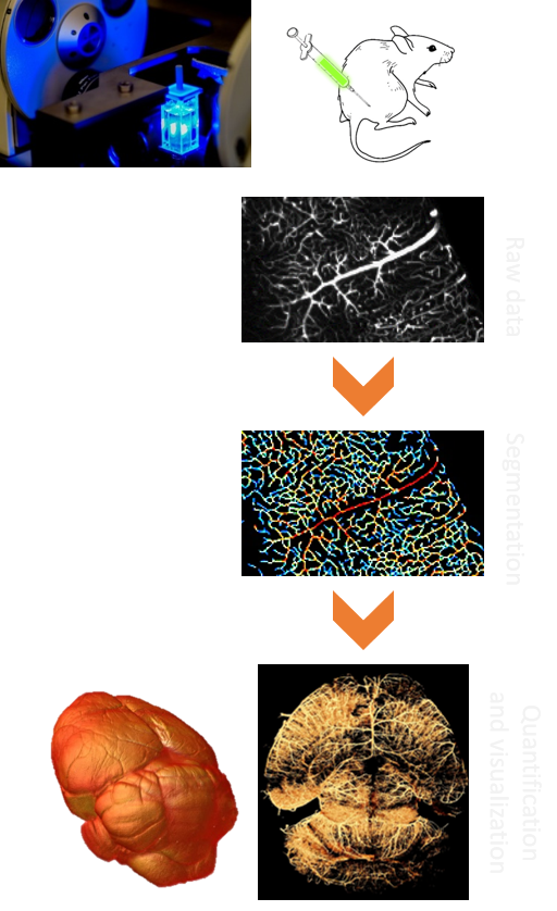
- Light sheet microscopy and clearing to characterize the vascular network in 3D
- Automated 3D image processing for micro-vascular network quantification
General Procedure
Prior to sample collection by Imactiv-3D:
- In vivo labelling by infusion with a fluorescent lectin before euthanasia
- Formalin fixation of extracted sample
Image acquisition:
- Optical clearing of samples
- 3D light sheet fluorescence microscopy
- Multi-position acquisition
Image processing and analysis:
- Quantitative characterization of the vascular network
- Vessels segmentation
- Extraction of efficient volume
- Computation of parameters of interest: vessels length and local size, density of the vascular network
- 3D visualization with surface and volume rendering
- Reconstruction of the whole sample
- Advanced display using 3D animations
Application example
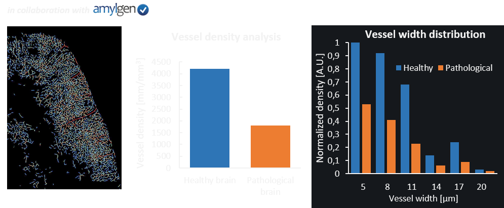
Study of Alzheimer pathology:
- Healthy brain versus pathology-induced brain.
- Diseased brain cortex shows a drastic decrease in vascular network density, together with a modified distribution of the vessels size.
- Results consistent with the inflammation induction described in the literature.